How will I take care of my pet following surgery?

Care of the incision:

You should monitor your pet’s incision at least once daily for evidence of infection. Infection generally appears as a painful red swelling associated with the incision. Other incisional problems include separation of the incision line. This is most commonly associated with infection or a pet licking or chewing at the incision. If you see your pet chewing at the incision he/she will need to wear an Elizabethan (a.k.a. lampshade) collar until the sutures or staples are taken out. Sutures are generally taken out around 2 weeks following the surgery.

Activity restriction:

Following surgery your animal will need to be confined to a crate or small room for 8 weeks. Slippery floors such as hardwood and ceramic tile should be avoided. Your pet should not be allowed to play with other animals or children and should be kept away from stairs. He or she should be taken outside 4 to 5 times per day to urinate and defecate. Your pet should be on a leash at all times when outside for at least 8 weeks.

Physical therapy:

Physical therapy following surgery for patellar luxation repair consists of massage and performing range of motion, as well as knowing when to start activities to encourage your pet to use the affected limb properly. Most dogs will place significant weight on the operated leg within 5-6 days of surgery. If your pet is not using the leg within 7-10 days post surgery then you should more closely follow the suggested range of motion exercises with him/her at least 3 times per day on the operated knee. Your veterinarian can instruct you about the specifics of physical therapy if indicated.

Recheck examinations:

Rechecks are generally scheduled for 2 weeks following surgery for suture removal and 6-8 weeks for final recheck examination. Xrays may be taken at specific intervals to evaluate healing. If your pet is doing well at that time no additional rechecks will be needed.

Medications:

Your veterinarian will provide you with instructions about medications to be taken following knee surgery. Generally some type of pain medication will be administered for 5 to 7 days following surgery.

General care:

Following any surgery you should also be sure that your pet is eating and drinking normally without vomiting, diarrhea or prolonged anorexia. You should also be aware of your pet’s general attitude and make sure that he or she interacts normally with your family members, is able to sleep normally and is not unusually lethargic. If you notice anything significantly out of the normal in reference to the above habits you should contact your veterinarian.

Postoperative Care

After surgery is completed, the affected leg(s) may be bandaged for three to seven days. Passive physical therapy is begun immediately after bandage removal to work out the stiffness and reestablish a normal range of motion in the joint. During the next three to four weeks, light walking around the house or supervised short walks outside must be strictly controlled until a progressive building of muscular support and stamina leads to unrestricted normal function.

Your pet must be kept in a confined area such as a small room, dog run, or kennel for the first 4 weeks following knee surgery. This confinement is essential to prevent your pet from damaging the repair caused by excessive activity such as running through the house and slipping on the tile/linoleum, when your pet sees a squirrel outside or when the doorbell rings. After this period of time, your pet needs to be kept on restricted activity for the next two months.

During this period of restricted activity he/she should still be confined to a small area, however slow, controlled leash walks are permitted. This restriction will allow sufficient time for the surgical site to heal completely. Tranquilizers may be necessary to help keep your pet quiet. Your pet may develop a tolerance to these drugs, however, necessitating progressively higher doses to effect the desired level of tranquilization. Only enough tranquilization to “take the edge off” for your pet and make them happy to walk rather than run is necessary. These drugs are not harmful or addictive to your pet in any way. No off-leash activity should be allowed for at least 10 weeks following surgery.

If a bandage is placed on your pet’s leg, it should be kept clean and dry. If there is a chance that the bandage will come in contact with water or moisture, you should cover the bandage with a plastic bag. Do not leave the plastic bag on your pet’s leg for a prolonged period of time however, or it will cause moisture accumulation and skin infection underneath the bandage! If your pet’s toes are exposed at the bottom of the bandage, please check them daily for any signs of swelling. If they should become cool and/or puffy, the bandage may be impeding circulation and should be changed immediately. If this should occur, please contact us.

If your pet’s toes are not exposed, pinch them through the bandage until your pet reacts to let you know he still has feeling in them. Due to your pet’s normal activity, the top may loosen and the incision site may become exposed. This is okay as long as your pet does not lick or chew at the incision area, the bandage still offers support to the knee, and you are still able to check your pet’s toes. If the incision area looks as though it needs to be cleaned, please clean the incision with witch hazel, and apply an antibiotic ointment to it if you wish.

If you find that your pet is having difficulty in rising and/or walking with the bandage on, we suggest that you use a towel placed around the stomach to act as a sling for the hind quarters. Lift your pet gently and support the rear limbs while it walks. We would be happy to show you how this is done. You can also purchase a premade sling from a pet store or online if you prefer.

Your pet may require a lot of patience and attention during this recovery period. Your biggest chore will be to keep your pet confined and inactive for a three month period. If you have any questions or if you need help and/or suggestions, please do not hesitate to contact our office.

Overall, some pets recover very quickly with little need for owners’ assistance, but other animals need intensive physical therapy. Lean and fit animals tend to recover quicker than overweight and weaker pets.

Exam, Screening Tests, and Imaging

The diagnosis of patellar luxation is essentially based on palpation of an unstable kneecap on orthopedic examination. Additional tests are usually required to diagnose conditions often associated with patellar luxation and help the surgeon recommend the most appropriate treatment for your pet.

These may include:

  • Palpation of the knee under sedation to assess damage to ligaments
  • Radiographs of the pelvis, knee and occasionally tibias to evaluate the shape of the bones in the rear leg and rule out hip dysplasia
  • 3-dimensional computed tomography to provide an image of the skeleton of the entire rear legs. This advanced imaging technique helps the surgeon plan surgery in cases where the shape of the femur or tibia needs to be corrected.

  • Because there is great individual variation in the pathologic deformities seen, a graded classification of medial patellar luxation (Putnam 1968) has been formulated as a basis for recommending which type of surgical repair is most appropriate for each individual.

    Surgical treatment is typically considered in grades 2 and over. While the grading system is useful in communicating the degree of patellar luxation, the anatomical abnormalities that might be present to produce the degree of luxation, which patients may require surgery at some point, and in suggesting the prognosis for surgical patients, there is a danger in reading too much into the classification system. For example, one cannot base recommendations for surgical repair solely on the grade of luxation present, because the correlation between the grade of luxation and the clinical signs is not strong.

    Many Grade 1 small dogs will never encounter lameness problems; however, others, especially many large breed dogs, will be clinically affected. On the other hand, nearly all Grade 3 and 4 dogs will show signs of lameness and disability. However, these signs are not always severe and, perhaps more importantly, some owners may not view the problem as significant in the context of the limited physical demands placed on their dogs, especially in the case of the small breeds.

    GRADE I

    The anatomic alignment of the stifle is normal with the patella luxating only when pushed out of the socket.

    GRADE II

    The patella is returned by manual pressure.

    GRADE III

    The patella is permanently dislocated but can be reduced manually with the limb extended.

    GRADE IV

    The patella is permanently dislocated and cannot be manually reduced. In dogs with grade IV patellar luxation more aggressive surgery is often required. This involves straightening of the femur and/or tibia with complete bone cuts (a.k.a. osteotomies) and stabilizing the bones with bone plates. Lateral patellar luxation, or LPL, is less common than MPL and occurs when the kneecap occasionally rides on the outside of its normal groove. It, too, can be congenital or acquired, with the congenital form again being more common. While it can occur in any dog, it is more common in large and giant breeds. LPL is frequently accompanied by malformation of the femur and/or tibia. The disease can produce marked lameness and progress to crippling arthritis. Because of the accompanying bony malformations, extensive surgery may be required to correct this problem.

What Options are Available for Treating Patellar Luxation?

Patellar luxations that do not cause any clinical sign should be monitored but do not typically warrant surgical correction, especially in small dogs. Surgery is considered in grades 2 and over. Surgical treatment of patellar luxation is more difficult in large breed dogs, especially when combined with cranial cruciate disease, hip dysplasia or angulation of the long bones. One or several of the following strategies may be required to correct patellar luxation: Reconstruction of soft tissues surrounding the knee cap to loosen the side toward which the patella is riding and tighten the opposite side.

Lateral Imbrication

This procedure alone may be adequate for a mild case but is often used as an adjunctive procedure to supplement one of the other surgeries. When the patella slips out of its groove, the joint capsule surrounding it is stretched to allow this motion. Imbrication simply involves taking a tuck in the joint capsule. The tightened joint capsule does not allow for the slipping of the kneecap and the kneecap is confined to its proper groove.

Trochleoplasty

Deepening of the trochlear groove, or trochleoplasty, can be accomplished with a variety of techniques. A chondroplasty technique involves cutting out a taco-shaped wedge of cartilage, removing a small portion of bone beneath it, and then replacing the cartilage. The result is a deeper groove. This procedure can only be performed on very young dogs, because their cartilage is thicker.

Trochlear recession involves cutting out the cartilage and bone in such a way as to create a deeper trough. This trough will then fill in with scar tissue over time. Because this scar tissue is not as good as cartilage for joint function, this technique has given way to others that attempt to preserve normal cartilage. It can, however, be useful in carefully selected cases.

Wedge recession creates a taco-shaped piece of cartilage and underlying bone. Then, the bone below the wedge is removed and the wedge is replaced, forming a deeper groove. Block recession is identical in principle to wedge recession, except that a rectangular piece of cartilage and bone, rather than a wedge, is removed.


Tibial Tuberosity Transposition

The kneecap attaches to the lower leg via its patellar tendon at a bony site called the tibial tuberosity. Many times this site forms abnormally on the inside, as with MPL, or on the outside, as with LPL. In this procedure, the surgeon fractures the tibial tuberosity and moves the tibial tuberosity back into proper alignment and secures it in place with a pin and or wire. Realigning the joint, kneecap, and tendon prevents dislocation from reoccurring.

Are there any potential complications associated with surgery?

Any surgical procedure has the potential for complications. Fortunately, surgery for a patellar luxation is commonly performed and complications are rare. Potential complications include problems with general anesthesia, surgical infection, migration of the surgical implants (i.e. pins that stabilize the tibial tuberosity transposition) associated with the repair, complete disruption of the tibial crest, rarely patellar tendon rupture and failure of the surgery to completely stabilize the patella and resolve lameness.

Greater than 90% of dogs with surgical repair of grade 1, 2 and 3 patellar luxations will not have any significant complications and will return to normal or near normal function with surgery. If pins had to be used to transpose the tibial crest, these may need to be removed in 3-12 months. Some dogs “allergic” to the metal. Ultimate results will depend on the amount of arthritis existing prior to surgery and/or the severity of the patellar luxation.

Incidence of Patellar Luxation

Patellar luxation is one of the most common congenital anomalies in dogs, diagnosed in 7% of puppies. The condition affects primarily small dogs, especially breeds such as Boston Terrier, Chihuahua, Pomeranian, Miniature Poodle and Yorkshire Terrier. The incidence in large breed dogs has been on the rise over the past ten years, and breeds such as Chinese Shar Pei, flat-coated retriever, Akita and Great Pyrenees are now considered predisposed to this disease. Patellar luxation affects both knees in 50% of all cases, resulting in discomfort and loss of function.

Intermittent or consistent lameness; bowlegged stance; reluctance to walk or jump; occasionally holding a rear leg out to the side when walking.

Lameness that is often intermittent, and may be unilateral or bilateral; thick, swollen stifles; pain on range-of-motion; crepitus; palpable luxation; inability to jump or walk normally; medial displacement of quadriceps muscle group; lateral bowing of the distal third of the femur.

Clinical signs associated with patellar luxation vary greatly with the severity of the disease: this condition may be an incidental finding detected by your veterinarian on a routine physical examination or may cause your pet to carry the affected limb up all the time. Most dogs affected by this disease will suddenly carry the limb up for a few steps, and may be seen shaking or extending the leg prior to regaining its full use.

As the disease progresses in duration and severity, this lameness becomes more frequent and eventually becomes continuous. In young puppies with severe medial patellar luxation, the rear legs often present a “bow-legged” appearance that worsens with growth. Large breed dogs with lateral patellar luxation may have a “knocked-in knee” appearance, combining severe lateral patellar luxation and hip dysplasia.


Causes of Patellar Luxation

Patellar luxation occasionally results from a traumatic injury to the knee, causing sudden non-weight-bearing lameness of the limb. It may also develop subsequent to cranial cruciate deficiency in dogs that will typically have a chronic history of lameness. However, the cause remains unclear in the majority of dogs. The femoral groove into which the kneecap normally rides is commonly shallow or absent in dogs with non–traumatic patellar luxation.

Early diagnosis of bilateral disease in the absence of trauma and breed predisposition supports the concept of patellar luxation resulting from a congenital or developmental misalignment of the entire extensor mechanism. Congenital patellar luxation is therefore no longer considered an isolated disease of the knee, but rather a component/consequence of a complex skeletal anomaly affecting the overall alignment of the limb, including:

  • Abnormal conformation of the hip joint, such as hip dysplasia
  • Malformation of the femur, with angulation and torsion
  • Malformation of the tibia
  • Deviation of the tibial crest, the bony prominence onto which the patella tendon attaches below the knee
  • Tightness/atrophy of the quadriceps muscles, acting as a bowstring
  • A patellar ligament that may be too long

Patellar Luxations

The patella, or kneecap, is a small bone buried in the tendon of the extensor muscles (the quadriceps muscles) of the thigh. The patella normally rides in a femoral groove within the stifle. The patellar tendon attaches on the tibial crest, a bony prominence located on the tibia, just below the knee. The quadriceps muscle, the patella and its tendon from the “extensor mechanism” and are normally well-aligned with each other. Patellar luxation is a condition where the knee cap rides outside the femoral groove when the stifle is flexed. It can be further characterized as medial or lateral, depending on whether the knee cap rides on the inner or on the outer aspect of the stifle.

When the patella is in its normal position, its cartilage surface glides smoothly and painlessly along the cartilage surface of the trochlear groove with little or no discomfort.

As the patella “pops out” of its groove these cartilage surfaces improperly rub each other. The animal may cry and try to straighten (extend) the leg to “pop it back in” or may hold the limb up until muscle relaxation allows the kneecap to reposition itself. This resembles an intermittent lameness.

There is little or no discomfort until the cartilage is effectively “rubbed off” or eroded to a point where bone touches bone. From this point on, each time the patella “pops out” into its abnormal, luxated position it will cause pain. This explains why many individuals have no clinical lameness until they reach adulthood. Often progressive cartilage wear creates an acutely painful condition.

Complications of Cruciate Surgery

Success Rate

Currently the success rate of either surgery is between 85-90%. This means your pet should get back to normal or near normal activity over a 2-4 month period. There are a small percentage of dogs and cats that do not do well following cruciate ligament injury, no matter how they are treated.

Anesthesia

There is always a risk with anesthesia although it is rare that significant problems are encountered if the pet has been thoroughly evaluated prior to surgery. All animals anesthetized are continuously monitored by a veterinary nurse throughout the procedure. They are placed on intravenous fluids and their heart rates, respiratory rates and blood pressure are constantly monitored. They are also placed on a circulating warm water blanket to help maintain their body temperature under anesthesia.

Infection

There is always the potential for infection with any surgery. In most cases infection occurs when the animal licks or chews at the surgical incision postoperatively. That is why we send your pet home with a protective collar that they should wear at all times to prevent them licking or chewing at the incision. With either surgery- if an infection develops it can delay healing and necessitate the removal of the implants (the stabilizing Nylon suture or the bone plate and screws), which is additional anesthesia and surgery for your pet and additional cost.

Implant failure

Premature breakdown of the stabilizing Nylon suture can occur and is more likely in very large, active dogs or obese dogs, which is why it is recommended that dogs over 70 pounds are suggested to have TTA procedures. It is also more likely if dogs do not have their activity restricted as directed postoperatively. If the Nylon suture breaks prematurely it can lengthen the recovery process and in some cases necessitate further surgery.

The bone plate and screws used in the TTA are very strong but by cutting the bone – this is essentially the same as having a fractured bone that requires time to heal. Initially the strength of the repair is provided by the plate and screws alone. Over the next few weeks, as the bone begins to heal, the bone starts providing additional strength to the repair. It is rare that we have implant failure with this technique but if the plate or screws break, the plate pulls off the bone, or the bone breaks around the screws further surgery is required and it can be very difficult to perform this additional repair. This is why it is crucial to follow the take home instructions for proper rest and rehabilitation.

Isolated meniscal injury

As previously stated, dogs do have a slight risk of developing an isolated meniscal injury in the future, requiring re-exploration of the joint. This can occur with either the lateral suture technique or the TTA and it is not possible to predict which dogs will be affected.

Fabella Pain

This is a potential complication of the lateral suture stabilization technique. The fabella is a small bone at the back of the dog’s knee that the Nylon stabilizing suture is placed around. In a very small percentage of dogs this can cause some discomfort and occasionally we need to remove it under a very short anesthesia.

Tibial Tuberosity fracture (fracture through the top, front part of the tibia or shin bone)

This is a rare complication of the TTA procedure. It occurs very infrequently and is most often secondary to the pet being too active postoperatively. It may require placement of metal pins to repair the damaged piece of bone.

Persistent Lameness secondary to osteoarthritis

This affects between 10-15% of dogs. This is a potential complication with either surgery and there is no way to predict which dogs will have significant problems with arthritic pain. It requires life-long activity moderation (less free running and jumping, more slow leash walks and swimming) and the use of non-steroidal anti-inflammatory drugs long term (drugs such as Rimadyl, Deramaxx, Metacam Galliprant and Previcox).

Specific instructions will be given at the time your pet is discharged from the hospital including what medications to give (antibiotics and pain medications). In general animals come back in 10-14 days to have their incisions checked and the skin sutures or staples removed. The stability and comfort of the joint will be checked at this visit. Following surgery your pet will require strict activity restriction. Patient activity is generally restricted for at least 2 ½ to 3 months.

Postoperative Instructions

For the first 10-14 days your dog should be confined to a crate or small room with minimal furniture. Steps should be kept to a minimum. They should go outside on a short leash, no longer than 3 feet (not at the end of an extendible leash) to go to the bathroom only – no free running, jumping or playing is allowed. This can result in failure of the implants and may result in the need for more surgery.

If your dog will tolerate it you can apply an ice pack to the incision up to 4 times daily for 10-20 minutes for the first 3-5 days. Do not try to force this if your dog will not let you. Once the skin sutures have been removed in 2 weeks you can start the following walking program at home – all other restrictions still apply and your dog should still not be allowed to roam free in the house: Week 3 post operatively: Slow, on a short leash walks for 5 minutes up to 3-4 times daily. You can also start making your dog walk in some small circles and figures of 8 with the operated leg on the inside of the circle/8.
Week 4 postoperatively: Slow, on a short leash walks for 10 minutes up to 3-4 times daily.
Week 5 postoperatively: Slow, on a short leash walks for 15 minutes up to 3-4 times daily.
Week 6 postoperatively: Slow, on a short leash walks for 20 minutes up to 3-4 times daily.

For dogs that have had a TTA, weeks 7 and 8 should be the same as week 6: Slow, on a short leash walks for 20 minutes up to 3-4 times daily.

If your dog is hard to control on the leash and is liable to injure themselves their activity should be completely restricted to leash walks to the bathroom only for the first 6 weeks. Following the lateral suture surgery dogs are then seen for a recheck at 6 weeks; further activity instructions will be given at this visit. For dogs that have had a TTA, follow-up x-rays will be needed to assess healing at 8 and 12 weeks postoperatively. There is an additional charge for these x-rays and sedation for at least the 8 week xrays,. Additional X-rays may be taken in 2 weeks if the 12 week radiographs do not show complete healing. Dogs that have had a lateral suture stabilization do not require follow-up x-rays in most cases.

All pets with stifle problems should maintain an ideal body weight and you may need to decrease the amount you are feeding during the postoperative recovery period.

The amount of arthritis that an animal develops after cruciate ligament injury is variable and there is no way to predict how it will affect your pet. It is important that once they have healed from surgery they have regular exercise, maintain an ideal body weight and, if possible, stay on a Hyaluronic Acid and Glucosamine/Chondroitin joint supplement for life.

In addition, some animals may require the tactical use of non-steroidal anti-inflammatory drugs such as Rimadyl, Deramaxx, Previcox Galiprant or Metacam. These can be obtained from your regular veterinarian and be used on an “as needed” basis. Arthritis tends to cause more discomfort after periods of sudden very heavy exercise (free running, jumping, ball chasing) and when the weather is cold and damp.

Bonnie Clarke

Bonnie joined us back in April of 2005 as a Veterinary Assistant. We refer to her as “BonBon” because she is one of the sweetest people you will ever meet. Bonnie has always been a great team player, ready and willing to lend a hand wherever she is needed.  At the start of her career with us, she loved working with all animals.  Her friendly and upbeat personality kept both clients and pets at ease throughout the appointment. She enjoyed working with the clients and patients during routine appointments, but had a special interest in ultrasound and x-ray.   These days Bonnie has moved into role of “General Overseer”. She keeps everything running smoothly by overseeing the inventory, keeping up with the laundry, and ensuring that the hospital is clean and sanitized.  With as busy as this hospital is we simply couldn’t manage without her. Family is very important to Bonnie so she works her schedule around assisting her parents and daughter. When not at work, Bonnie loves spending time with her daughter and her dogs, Patrick and Ginger. She also enjoys swimming, tanning and yard work.  Clients and staff simply adore Bonnie. We are so lucky to have her here.

Nancy Graver

Nancy has been a part of this family since May of 2005 as a part-time receptionist/patient coordinator. The clients love Nancy. She makes a point of knowing the clients and their pets by name and keeps up on what’s going on in their lives. She has a way of making everyone feel welcome and right at home. She came to us after retiring from Verizon after 33 years, during which time she won many customer service awards. Nancy has recently moved up to full-time hours. Nancy’s favorite part of this job is the opportunity she gets to interact with all of the great pets and clients we see here at the Back Mountain Veterinary Hospital. Children who come to the appointments with their parents are treated to stickers and lollipops that Nancy always keeps in stock. Nancy is a favorite amongst the staff here as well. She spoils us with homemade cupcakes and surprises us on occasion with great fruit salad and trays of kielbasa and cheese. When she is not making everyone around her feel special, she enjoys antiquing and decorating her home and garden with all of her unique “finds.”

Gina Masters

Gina joined the ranks in October of 2010 as a part-time receptionist/patient coordinator. She quickly moved up to full-time, bringing her “high energy” work style into full gear. Gina is the life of the party, and usually found at the center of things whenever laughter is heard ringing through the halls. However, she takes getting to know the clients and patients very seriously. That drive to provide great customer service has helped her advance into her current role, Patient Care Supervisor, where she oversees the insane details necessary to keep the Front Office running smoothly.  Even after all these years, Gina still strives to learn something new every day and making clients feel at home is always her top priority. Speaking of home, Gina has a son, Logan, with special needs and so has co-founded the L.O.G.A.N. Foundation in his honor – which stands for Little Ones Genetic and Neurological. She also coaches gymnastics and special needs cheerleading, as the owner of Masters Athletic Elite.  She manages all-star gym and tumble teams, as well as being head coach for DHS varsity cheerleading. Family means the world to Gina and she enjoys the time she gets to spend with her husband, son, daughter Cassandra and Vito, the big family Newfie.

Joy Mueller

Joy is the equivalent of the family matriarch, having been a part of the Back Mountain Veterinary Hospital over 15 years. She started as a Veterinary Assistant and often pitched in as a Receptionist/Patient Coordinator when needed. Joy worked her way up to Surgical Technician, then after several years of teching, she took over as Hospital Administrator. Although she misses the hands-on care of the patients, she finds working behind the scenes to keep the hospital running at peak performance just as rewarding. Luckily, she gets to keep her Tech skills sharp by pitching in when we are short-handed and by being the “go-to” person when there is a difficult blood draw or catheter placement. Clients let her know how much she is missed, but they appreciate having someone whom they have come to know and trust always available if they have a question or concern. The staff likes knowing Joy understands the demands of their job and is always ready to lend a hand on busy days. What Joy likes best about working here is being a part of the constant growth and advancements Dr. Phillips strives for to ensure that his hospital always provides the best possible care.

Kim Petros, LVT

Kim came onboard with us in July of 2009 as a Veterinary Technician, after getting her Associate Degree in Veterinary Science from Johnson College. Kim has grown into a great addition to our staff. She is personable and dependable, always willing to take on the extra responsibility of managing our overnight intensive care patients. Kim is a Licensed Veterinary Technician, getting her State Certification in August 2016. Her Certification has allowed her to become a mentor and trainer for the next generation of Johnson College students. Kim enjoys just about everything about the job, but has a special interest in veterinary dentistry and is gaining valuable skills in dental radiology. She also certified to provide Therapeutic Laser Treatments. She has a special love for working with puppies and kittens, educating owners to ensure that they get off to a good start.  She is taking on more leadership here in her role as Head Veterinary Technician and takes pride in being able to put her many advanced skills to good use in a hospital that values quality, comprehensive, state-of-the-art care for its patients. Kim manages the stress of the veterinary field with her love of reading and physical fitness.  She is a fanatic about Halloween and Christmas, playing only Christmas music in surgery from the very beginning of November. She treats her cat, Dinah, like a princess, even getting her portrait painted in regal attire. In her free time, Kim enjoys shopping and traveling and hopes to someday go on Safari in Africa.  For now she’ll just settle for visiting local breweries and wineries.

Mia Remplewicz

Mia became a valued member of the BMVH family on April 1st, 2019, which should have been our warning to just how big a practical joker she was going to be!  Like the time she covered brussels sprouts in white chocolate and sprinkles and told the staff they were cake pops! The staff’s reactions were pretty priceless!  Mia’s great sense of humor, even if its at her own expense, really helps all of us get through the stressful and sad times that are unfortunately unavoidable in veterinary medicine.  We don’t ever want to know what its like to be without her wit, her willingness to break into song, or her ability to quote from her favorite tv show, The Office. Equally important, Mia is an excellent technician, with a keen interest in anesthesia and surgery.  She especially enjoys scrubbing in to assist for difficult or complex surgeries.  Mia is excited by the constant advancements in Veterinary Medicine and loves the challenge of always learning something new. She is patient, detail-oriented and very caring, qualities which extend into her love of training new staff to ensure their success in Veterinary Medicine. Clients really appreciate the compassion and empathy she brings to the exam rooms, along with her willingness to answer questions and help educate clients so they can take to best possible care of their fur-babies. Speaking of fur-babies, Mia loves to spend time with her two dogs, Wyatt the Great Dane and Leroy the Silver Lab.  She and her fiance, Joey, love spending time outdoors. They are always up for an adventure, be it 4-wheeling in a mud bog, camping under the stars or simply taking care of their chickens and ducks, oh yeah, and can’t forget the hamster, Norman Bates! We told you she had a great sense of humor!

Dr. Brock Phillips

Dr. Brock Phillips is the Chief Veterinarian and owner of the Back Mountain Veterinary Hospital. Dr. Phillips has been a practicing small animal veterinarian and a part of this community for over 30 years. After completing his undergrad studies at Ithaca College, he went on to Araneta University, College of Veterinary Medicine. Dr. Phillips completed his externship at the University of Tennessee, College of Veterinary Medicine. Dr. Phillips loves to learn and grow as Veterinary Medicine evolves. Through the years he has taken extensive Continuing Education in Laser Surgery, Ultrasound, Echocardiograms, Endoscopy and Cold Laser Therapy. He has been a leader in Luzerne County for bringing quality advanced diagnostic and surgical capabilities into the day practice setting. Dr. Phillip’s forethought and guidance has made the Back Mountain Veterinary Hospital an exceptional Referral Center for the surrounding areas as well as a day practice the local community has come to rely upon. Our new hospital has been a labor of love over many long years.  Dr. Phillips personally supervised each and every element from the layout of the floor plan to the decor and color scheme, ensuring the hospital has the optimal flow for maximizing patient care, while providing a calm, comfortable atmosphere to minimize stress for anxious pet parents. In what little free time he can call his own, Dr. Phillips enjoy cycling, hiking, and tennis. He rarely gets to vacation, but when he can he loves to snorkel and scuba dive. Dr. Phillips has a background in music and played the trombone in college. He is greeted each evening by his furry friend, Dewey the Cat!

Dr. Kelly Brooks

Dr. Kelly Brooks joined the Back Mountain Veterinary Hospital in March of 2012. She brings a fresh perspective and a lot of energy to our team. Dr. Brooks did her undergrad and grad school studies at the University of Arizona, with her Bachelor’s and Master’s Degrees in Physiology. She then went on to pursue her studies at The Ohio State University, College of Veterinary Medicine. After graduating, she completed an internship at the Advanced Veterinary Care Center in California by doing a 1 year rotation of small animal medicine and surgery.   Dr. Brooks is an active member of the American Veterinary Medical Association and the Pennsylvania Veterinary Medical Association. She has developed quite a skilled with soft tissue surgery and has come a trusted resource for certain eye surgeries, including Grid Keratotomy, Cherry Eye, and Enucleation. She has a great love for Cardiology (pun intended) and a special interest in Internal Medicine, focusing on Endocrinology, Gastroenterology, Hematology and Blood Disorders.  These interests make her very pleased to be a part of a facility with a long-standing history as a Referral Center for Advanced Diagnostics, offering Echocardiograms, Abdominal Ultrasounds, and Digital Radiology with consults from Cardiology, Internal Medicine, and Radiology Specialists.   When not hard at work, Dr. Brooks enjoys the physical stress release of running and hiking, as well as the mental rejuvenation that comes from photography, painting, and yoga. She especially enjoys goat yoga! When the weather isn’t conducive to her outdoor activities, Dr. Brooks either indulges her passion for finding vintage treasures at the Salvation Army and local antiques shops, or she puts her incredible green thumb to work on her in-home plant jungle for her cats Amelia and Mac.

Dr. Amy Morris

Dr. Amy Morris became a part of the Back Mountain family in April of 2017. After completing her undergrad studies at the Bryn Mawr College, and completing an internship at the Cincinnati Zoo, she went on to study at Ohio State College of Veterinary Medicine. Dr. Morris did a 2-week externship at Best Friends Animal Sanctuary during vet school, then went straight into medical practice after graduating and obtaining her Veterinary License in 2010. She worked for 3 other Veterinary Hospitals before joining us, where she gained valuable experience in small animal and exotic pet medicine. Her work as a solo practitioner really helped to give her confidence to think calmly and perform well under pressure. Dr. Morris has been a member of the AVMA since a student in 2006. She has won an award for her Public Health Project, “Paws a Moment — Think Safety!” which uses dogs otherwise at risk of developing behavior issues to find a purpose by helping teach children about safety issues.  This included lessons on safe interactions with dogs and bite prevention. She is a Category II USDA Certified Veterinarian. Dr. Morris has a special interest Integrative Medicine, including certification as a Level II practitioner of Healing Touch for Animals. Client education is very important to Dr. Morris to optimize pet wellness and to prevent the spread of disease.  She is an advocate for Positive Behavior and encourages pet owners to bring nervous pets in for simple “happy visits” to alleviate anxiety and stress for both the pet and pet parents about vet appointments. Dr. Morris also worked in a neurology research lab before attending vet school, which really sparked her love for all things Neurology related!  She is very happy to be a part of the BMVH team and enjoys helping us grow, espeically in the field of medicinal and surgical care for Exotics. She is even the driving force behind the hospital’s strive to get the RHDV2 vaccine to protect rabbits from this terrible disease. When not hard at work, she also enjoys hiking with her dog, Elijah and spending time enriching the lives of her cats, Black Jack, MeLaan and Crowley.   She is excited to have a work schedule that will allow her to spend quality time with her Fiance, Chris, and to indulge in some baking for family and friends.  The staff get treated to personal containers of her cookies every year!

Dr. Anjilla Cooley

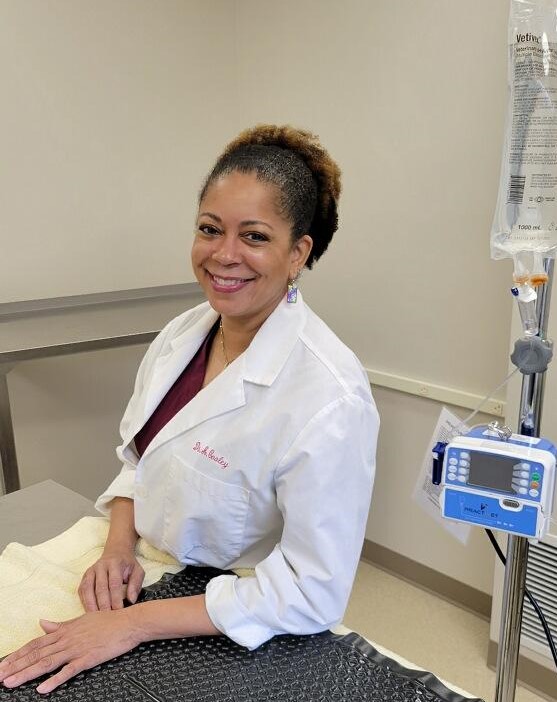
Dr. Anjilla Cooley has been coming to the Back Mountain Veterinary Hospital for the last 15 years to perform orthopedic surgery, such as ACL repair and compound fracture plating, as well as advanced soft tissue surgery and procedures like Myelograms and complex tumor resections. Dr. Cooley is available at Back Mountain Veterinary Hospital on a weekly basis to address your surgical needs. Dr. Cooley’s educational background consists of receiving a Doctorate of Veterinary Medicine from the University of Tennessee in 1992. In 1993, a certificate of completion for Small Animal Medicine and Surgery Internship was obtained from the University of Missouri in Columbia, MO. She completed her Small Animal Surgical Residency and obtained a Master’s Degree in Veterinary Science from Virginia-Maryland Regional College of Veterinary Medicine in Blacksburg, VA during the years of 1993 thru 1996. Dr. Cooley also taught as a lecturer at The Cornell University Veterinary Teaching Hospital in Ithaca, NY from 1996 thru 1999. Consequently, she created and implemented a didactic course at Cornell University, which became part of the required curriculum. Her employment tenure includes Round Valley Referral Hospital in Lebanon, NJ from 1999 thru 2001, Valley Central Referral Hospital in Whitehall, PA from 2001 thru 2005, and Crown Veterinary Specialists, in Lebanon NJ from 2005 thru 2011 where she was employed as a staff surgeon. In conjunction, she has traveled throughout Pennsylvania and New Jersey performing numerous advanced orthopedic, neurologic and soft tissue surgeries at various hospitals and clinics since 1999.